Book Easily
Customer support 24/7 throughout the week

Comprehensive full-body skin examinations, dermoscopic mole evaluations, biopsies, and on-site minor surgical removals at Panorama GP Medical Hub
Get In TouchComprehensive full-body skin examinations, dermoscopic mole evaluations, biopsies, and on-site minor surgical removals at Panorama GP Medical Hub.
Systematic head-to-toe clinical evaluation of moles, spots, and skin lesions using high-magnification polarising dermoscopy.
Punch and shave biopsies performed under local anesthesia to obtain tissue samples for immediate histopathological analysis.
On-site surgical removal of confirmed melanomas, basal cell carcinomas (BCC), and squamous cell carcinomas (SCC) with clear margins.
Liquid nitrogen cryosurgery for non-invasive pre-cancerous lesions, solar keratoses (sun spots), and superficial skin dysplasias.
Photographic recording and monitoring of high-risk dysplastic naevi to detect subtle structural or color changes over time.
Suture removal, clinical wound dressing changes, scar management advice, and histological result discussions.
The most serious form of skin cancer, originating in pigment-producing melanocytes. Early detection through dermoscopy is critical to prevent deep tissue invasion and metastasis.
The most common non-melanoma skin cancer. BCC grows slowly and rarely spreads, appearing as a pearly nodule, firm red spot, or non-healing sore.
A non-melanoma cancer developing in squamous cells. Appears as a scaly, tender, or thickened red patch that can bleed or crust over time.
Patients should inspect their skin monthly. Book a clinical check immediately if any spot exhibits the following features:
Individual risk increases significantly for patients meeting specific clinical profiles:
Doctors utilize handheld dermoscopes to evaluate subsurface skin structures without invasive initial procedures.
Biopsies and surgical excisions are performed on-site using sterile equipment under local anesthesia.
Excised tissue samples are sent directly to accredited laboratories for formal pathology examination.
Visual inspection of skin surfaces using dermoscopy, recording high-risk lesions and patient history.
Immediate cryotherapy for solar keratoses or scheduled biopsy for suspicious lesions requiring analysis.
Laboratory testing of samples followed by surgical excision if malignancy is confirmed by histology.
Post-operative wound inspection, suture removal, scar mitigation advice, and future monitoring plans.
To ensure maximum clinical accuracy during your full-body skin assessment, please follow these instructions:
A thorough skin check takes approximately 15 to 20 minutes, during which the doctor systematically inspects all skin areas, including scalp, ears, back, and limbs.
Adults in high UV environments like Western Australia should undergo a professional skin check annually. High-risk patients with a history of melanoma may require checks every 3 to 6 months.
Skin biopsies and excisions are performed under local anesthesia. You may feel a brief sting during the numbing injection, but the procedure itself is pain-free.
Pathology results generally return from the laboratory within 3 to 7 working days. A follow-up appointment will be scheduled to discuss the findings and determine if further surgical action is necessary.
Medicare rebates apply to medically necessary skin checks, diagnostic biopsies, and surgical excisions. Specific out-of-pocket costs depend on the complexity and location of the procedure.
What to Expect During Your Consultation
Initial Assessment
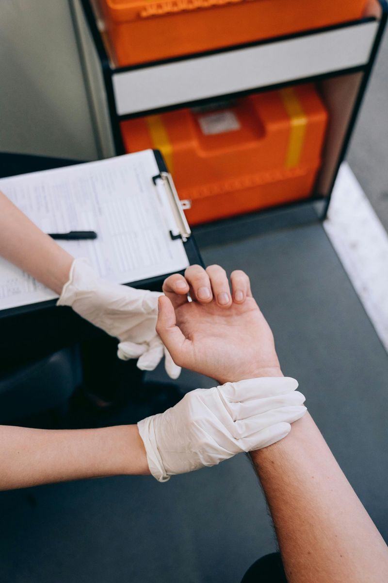
Review of current symptoms, medical history, active medications, and relevant lifestyle risk factors.
Clinical Examination
Physical examination, vital sign checks, or targeted diagnostic testing based on presenting symptoms.
Treatment Plan
Issuance of necessary prescriptions, diagnostic imaging requests, pathology referrals, or specialist advice.
Follow-Up Scheduling
Arrangement of subsequent appointments to review test results, monitor treatment effectiveness, or adjust medication plans.